Identification of a Brainstem Circuit Regulating Visual Cortical State in Parallel with Locomotion

Sensory processing is dependent upon behavioral state. In mice, locomotion is accompanied by changes in cortical state and enhanced visual re- sponses. Although recent studies have begun to elucidate intrinsic cortical mechanisms underlying this effect, the neural circuits that initially couple locomotion to cortical processing are unknown. The mesencephalic locomotor region (MLR) has been shown to be capable of initiating running and is associated with the ascending reticular activating system. Here, we find that optogenetic stimulation of the MLR in awake, head-fixed mice can induce both locomotion and increases in the gain of cortical responses. MLR stimulation below the threshold for overt movement similarly changed cortical pro- cessing, revealing that MLR’s effects on cortex are dissociable from locomotion. Likewise, stimulation of MLR projections to the basal forebrain also enhanced cortical responses, suggesting a pathway linking the MLR to cortex. These studies demon- strate that the MLR regulates cortical state in parallel with locomotion.

A. Moses Lee, Jennifer L. Hoy, Antonello Bonci, Linda Wilbrecht, Michael P. Stryker, and Cristopher M. Niell, Identification of a Brainstem Circuit Regulating Visual Cortical State in Parallel with Locomotion, Neuron 83, 455–466, July 16, 2014, http://dx.doi.org/10.1016/j.neuron.2014.06.031

Identification of a Brainstem Circuit Regulating Visual Cortical State in Parallel with Locomotion2014-07-16T21:45:00+00:00

Cocaine-induced structural plasticity in frontal cortex correlates with conditioned place preference

Contextual cues associated with previous drug exposure can trigger drug craving and seeking, and form a substantial obstacle in substance use recovery. Using in vivo imaging in mice, we found that cocaine administration induced a rapid increase in the formation and accumulation of new dendritic spines, and that measures of new persistent spine gain correlated with cocaine conditioned place preference. Our data suggest that new persistent spine formation in the frontal cortex may be involved in stimulant-related learning driving appetitive behavior.

Francisco Javier Muñoz-Cuevas, Jegath Athilingam, Denise Piscopo, Linda Wilbrecht, Cocaine-induced structural plasticity in frontal cortex correlates with conditioned place preference, Nature Neuroscience (2013) doi:10.1038/nn.3498 (Full Text)

Cocaine-induced structural plasticity in frontal cortex correlates with conditioned place preference2013-08-25T16:33:52+00:00

Transient stimulation of distinct subpopulations of striatal neurons mimics changes in action value

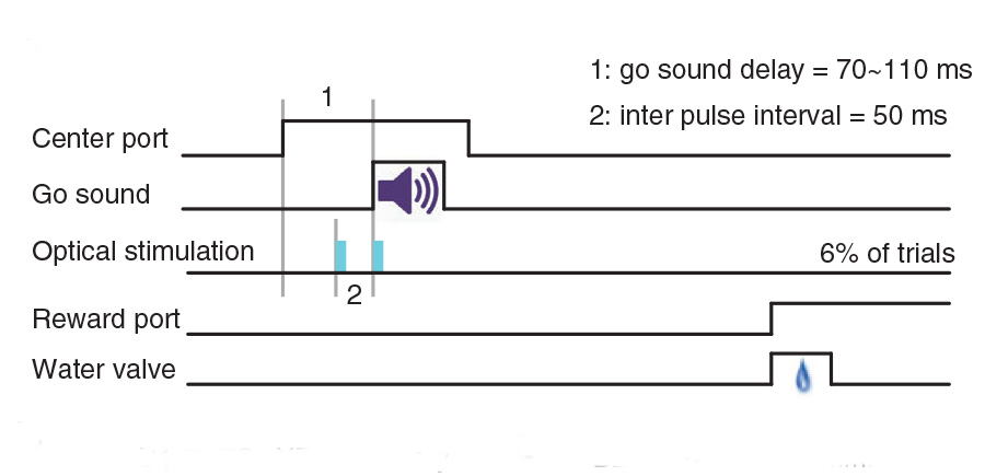
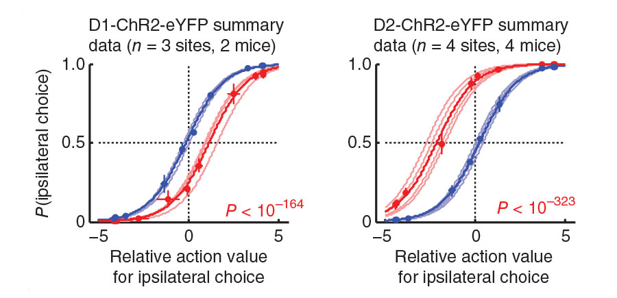
In changing environments, animals must adaptively select actions to achieve their goals. In tasks involving goal-directed action selection, striatal neural activity has been shown to represent the value of competing actions. Striatal representations of action value could potentially bias responses toward actions of higher value. However, no study to date has demonstrated the direct effect of distinct striatal pathways in goal-directed action selection. We found that transient optogenetic stimulation of dorsal striatal dopamine D1 and D2 receptor–expressing neurons during decision-making in mice introduced opposing biases in the distribution of choices. The effect of stimulation on choice was dependent on recent reward history and mimicked an additive change in the action value. Although stimulation before and during movement initiation produced a robust bias in choice behavior, this bias was substantially diminished when stimulation was delayed after response initiation. Together, our data suggest that striatal activity is involved in goal-directed action selection.

Lung-Hao Tai*, A Moses Lee*, Nora Benavidez, Antonello Bonci, Linda Wilbrecht, Transient stimulation of distinct subpopulations of striatal neurons mimics changes in action value Nature Neuroscience 15, 1281–1289, (2012) doi:10.1038/nn.3188 (Full Text)

Transient stimulation of distinct subpopulations of striatal neurons mimics changes in action value2012-08-19T16:51:22+00:00

Juvenile mice show greater flexibility in multiple choice reversal learning than adults

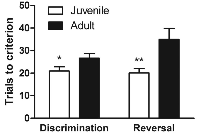
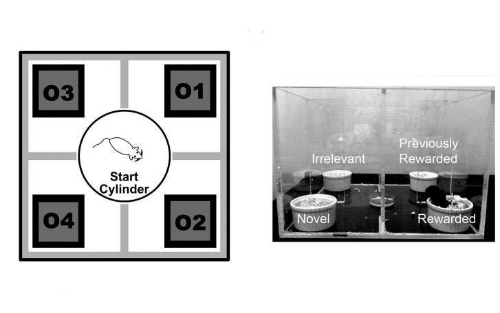
We hypothesized that decision-making strategies in juvenile animals, rather than being immature, are optimized to navigate the uncertainty and instability likely to be encountered in the environment at the time of the animal’s transition to independence. We tested juvenile and young adult mice on discrimination and reversal of a 4-choice and 2-choice odor-based foraging task. Juvenile mice (P26–27) learned a 4-choice discrimination and reversal faster than adults (P60–70), making fewer perseverative and distraction errors. Juvenile mice had shorter choice latencies and more focused search strategies. In both ages, performance of the task was significantly impaired by a lesion of the dorsomedial frontal cortex. Our data show that the frontal cortex can support highly flexible behavior in juvenile mice at a time coincident with weaning and first independence. The unexpected developmental decline in flexibility of behavior one month later suggests that frontal cortex based executive function may not inevitably become more flexible with age, but rather may be developmentally tuned to optimize exploratory and exploitative behavior for each life stage.

Johnson C and Wilbrecht L. 2011. Juvenile mice show greater flexibility in multiple choice reversal learning than adults. Dev Cogn Neurosci. 2011 Oct;1(4):540-51. doi: 10.1016/j.dcn.2011.05.008 (Full Text)

Juvenile mice show greater flexibility in multiple choice reversal learning than adults2011-10-01T11:35:21+00:00

Structural Plasticity Underlies Experience-Dependent Functional Plasticity of Cortical Circuits

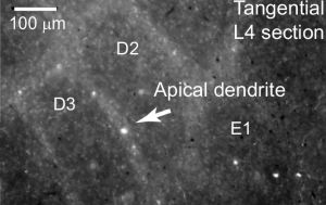
The stabilization of new spines in the barrel cortex is enhanced after whisker trimming, but its relationship to experience-dependent plasticity is unclear. Here we show that in wild-type mice, whisker potentiation and spine stabilization are most pronounced for layer 5 neurons at the border between spared and deprived barrel columns. In homozygote αCaMKII-T286A mice, which lack experience-dependent potentiation of responses to spared whiskers, there is no increase in new spine stabilization at the border between barrel columns after whisker trimming. Our data provide a causal link between new spine synapses and plasticity of adult cortical circuits and suggest that αCaMKII autophosphorylation plays a role in the stabilization but not formation of new spines.

Wilbrecht L, Holtmaat A, Wright N, Fox K, Svoboda K. 2010. Structural plasticity supports experience-dependent functional plasticity of cortical circuits. The Journal of Neuroscience, 7 April 2010, 30(14): 4927-4932; doi: 10.1523/​JNEUROSCI.6403-09.2010 (Full Text)

Structural Plasticity Underlies Experience-Dependent Functional Plasticity of Cortical Circuits2010-04-07T14:17:17+00:00

Neural circuits can bridge systems and cognitive neuroscience

There has been an emerging focus in neuroscience research on circuit-level interaction between multiple brain regions and behavior. This broad circuit-level approach creates a unique opportunity for convergence and collaboration between studies of humans and animal models of cognition.

Wilbrecht L, Shohamy D. 2010. Neural circuits can bridge systems and cognitive neuroscience. Front Hum Neurosci. 3:81 doi: 10.3389/neuro.09.081.2009 (Full Text)

Neural circuits can bridge systems and cognitive neuroscience2010-01-20T14:05:16+00:00

Long-term, high-resolution imaging in the mouse neocortex through a chronic cranial window

To understand the cellular and circuit mechanisms of experience-dependent plasticity, neurons and their synapses need to be studied in the intact brain over extended periods of time. Two-photon excitation laser scanning microscopy (2PLSM), together with expression of fluorescent proteins, enables high-resolution imaging of neuronal structure in vivo. In this protocol we describe a chronic cranial window to obtain optical access to the mouse cerebral cortex for long-term imaging. A small bone flap is replaced with a coverglass, which is permanently sealed in place with dental acrylic, providing a clear imaging window with a large field of view (~0.8–12 mm2). The surgical procedure can be completed within ~1 h. The preparation allows imaging over time periods of months with arbitrary imaging intervals. The large size of the imaging window facilitates imaging of ongoing structural plasticity of small neuronal structures in mice, with low densities of labeled neurons. The entire dendritic and axonal arbor of individual neurons can be reconstructed.

Holtmaat A, Bonhoeffer T, Chow, Chuckowree J, De Paola V, Hofer S, Hubener M, Keck T, Lee W-C A, Knott G, Mrsic-Flogel T, Mostany R, Nedivi E, Portera-Cailliau C, Svoboda K, Trachtenberg J, Wilbrecht L. 2009. Long-term, high-resolution imaging in the mouse neocortex through an imaging window. Nature Protocols 4(8):1128-44. (Full Text)

Long-term, high-resolution imaging in the mouse neocortex through a chronic cranial window2009-07-16T14:45:49+00:00

Linking Affect to Action: Critical Contributions of the Orbitofrontal Cortex

The orbitofrontal cortex (OFC) is a brain region that is privy to a wealth of information from sensory, emotional, and memory-related brain regions and thus likely serves as an important center for integration and evaluation. Recent research is revealing that the OFC could be a major site, or is at the very least an essential participant in a network of sites, where sensory and memory-related information is evaluated and transformed into predictions of the future used to guide decisions and actions.

On March 11–14, 2007, the New York Academy of Sciences hosted a conference dedicated to integrating recent research on the orbitofrontal cortex. The overarching goal of the event was to better define the function of the OFC. The conference included sessions on the effects of OFC damage on behavior, encoding value in the OFC, updating associations in the OFC, OFC anatomy and function, and translating OFC research. The proceedings of this conference were gathered in a volume of the Annals of the New York Academy of Sciences.

Wilbrecht L. 2007. Linking Affect to Action: Critical Contributions of the Orbitofrontal Cortex. eBriefing on a New York Academy of Science March 2007 conference.

Linking Affect to Action: Critical Contributions of the Orbitofrontal Cortex2007-03-01T15:01:16+00:00

High levels of new neuron addition persist when the sensitive period for song learning is experimentally prolonged

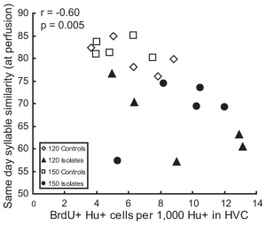
Socially reared zebra finch males imitate a song they hear during posthatching days 30–65; during this time, many new neurons are added to the high vocal center (HVC), a forebrain nucleus necessary for the production of learned song. New neuron addition drops sharply after day 65, and no new songs are imitated. In contrast, male zebra finches reared in isolation from other males have more variable songs at day 65 and thereafter can still imitate new sounds (Eales, 1985). We show that, in isolate birds, a greater number of new neurons continues to be added to HVC during the next 85 d, and this number correlates with syllable variability. We suggest that new neuron addition and turnover facilitate song change and that this effect lingers when an expected learning event is delayed.

Key words: neurogenesis; learning and memory; BrdU; birdsong; neuroethology; deprivation

Wilbrecht L, Williams H, Gangadhar N, Nottebohm F. 2006. High levels of new neuron addition persist when the sensitive period for song learning is experimentally prolonged. J Neurosci. 26(36):9135-41. (Full Text)

High levels of new neuron addition persist when the sensitive period for song learning is experimentally prolonged2006-09-06T15:21:53+00:00

Spine growth precedes synapse formation in the adult neocortex in vivo

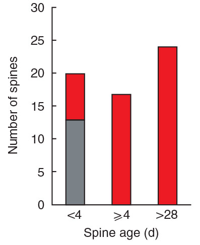
Dendritic spines appear and disappear in an experience-dependent manner. Although some new spines have been shown to contain synapses, little is known about the relationship between spine addition and synapse formation, the relative time course of these events, or whether they are coupled to de novo growth of axonal boutons. We imaged dendrites in barrel cortex of adult mice over 1 month, tracking gains and losses of spines. Using serial section electron microscopy, we analyzed the ultrastructure of spines and associated boutons. Spines reconstructed shortly after they appeared often lacked synapses, whereas spines that persisted for 4 d or more always had synapses. New spines had a large surface-to-volume ratio and preferentially contacted boutons with other synapses. In some instances, two new spines contacted the same axon. Our data show that spine growth precedes synapse formation and that new synapses form preferentially onto existing boutons.

Knott GW, Holtmaat A, Wilbrecht L, Welker E, Svoboda K. 2006. Spine growth precedes synapse formation in the adult neocortex in vivo. Nature Neuroscience 9, 1117 – 1124 (2006)
Published online: 6 August 2006 | doi:10.1038/nn1747 (Full Text)

Spine growth precedes synapse formation in the adult neocortex in vivo2006-08-06T15:40:06+00:00