Tag: Patricia Janak
-
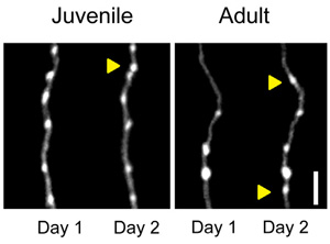
Long-range orbitofrontal and amygdala axons show divergent patterns of maturation in the frontal cortex across adolescence
The adolescent transition from juvenile to adult is marked by anatomical and functional remodeling of brain networks. Currently, the cellular and synaptic level changes underlying the adolescent transition are only coarsely understood. Here, we use two-photon imaging to make time-lapse observations of long-range axons that innervate the frontal cortex in the living brain. We labeled…