Tag: Karel Svoboda
-
Structural Plasticity Underlies Experience-Dependent Functional Plasticity of Cortical Circuits
The stabilization of new spines in the barrel cortex is enhanced after whisker trimming, but its relationship to experience-dependent plasticity is unclear. Here we show that in wild-type mice, whisker potentiation and spine stabilization are most pronounced for layer 5 neurons at the border between spared and deprived barrel columns. In homozygote αCaMKII-T286A mice, which…
-

Long-term, high-resolution imaging in the mouse neocortex through a chronic cranial window
To understand the cellular and circuit mechanisms of experience-dependent plasticity, neurons and their synapses need to be studied in the intact brain over extended periods of time. Two-photon excitation laser scanning microscopy (2PLSM), together with expression of fluorescent proteins, enables high-resolution imaging of neuronal structure in vivo. In this protocol we describe a chronic cranial…
-
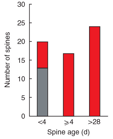
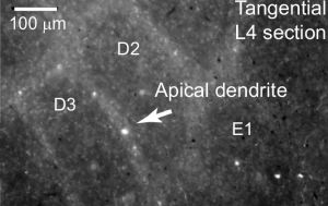
Spine growth precedes synapse formation in the adult neocortex in vivo
Dendritic spines appear and disappear in an experience-dependent manner. Although some new spines have been shown to contain synapses, little is known about the relationship between spine addition and synapse formation, the relative time course of these events, or whether they are coupled to de novo growth of axonal boutons. We imaged dendrites in barrel…
-

Experience-dependent and cell-type specific spine growth in the neocortex
Functional circuits in the adult neocortex adjust to novel sensory experience, but the underlying synaptic mechanisms remain unknown. Growth and retraction of dendritic spines with synapse formation and elimination could change brain circuits. In the apical tufts of layer 5B (L5B) pyramidal neurons in the mouse barrel cortex, a subset of dendritic spines appear and…
-
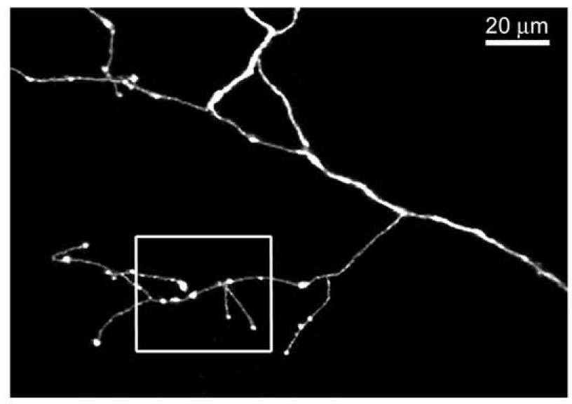
Cell type-specific structural plasticity of axonal branches and boutons in the adult neocortex
We imaged axons in layer (L) 1 of the mouse barrel cortex in vivo. Axons from thalamus and L2/3/5, or L6 pyramidal cells were identified based on their distinct morphologies. Their branching patterns and sizes were stable over times of months. However, axonal branches and boutons displayed cell type-specific rearrangements. Structural plasticity in thalamocortical afferents…
-
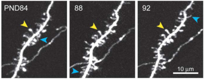
Transient and Persistent Dendritic Spines in the Neocortex In Vivo
Dendritic spines were imaged over days to months in the apical tufts of neocortical pyramidal neurons (layers 5 and 2/3) in vivo. A fraction of thin spines appeared and disappeared over a few days, while most thick spines persisted for months. In the somatosensory cortex, from postnatal day (PND) 16 to PND 25 spine retractions…