Tag: Gordon M. Shepherd
-
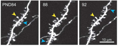
Transient and Persistent Dendritic Spines in the Neocortex In Vivo
Dendritic spines were imaged over days to months in the apical tufts of neocortical pyramidal neurons (layers 5 and 2/3) in vivo. A fraction of thin spines appeared and disappeared over a few days, while most thick spines persisted for months. In the somatosensory cortex, from postnatal day (PND) 16 to PND 25 spine retractions…