Tag: A. Wren Thomas
-
Does puberty mark a transition in sensitive periods for plasticity in the associative neocortex?
Postnatal brain development is studded with sensitive periods during which experience dependent plasticity is enhanced. This enables rapid learning from environmental inputs and reorganization of cortical circuits that matches behavior with environmental contingencies. Significant headway has been achieved in characterizing and understanding sensitive period biology in primary sensory cortices, but relatively little is known about…
-
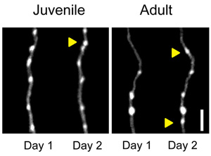
Long-range orbitofrontal and amygdala axons show divergent patterns of maturation in the frontal cortex across adolescence
The adolescent transition from juvenile to adult is marked by anatomical and functional remodeling of brain networks. Currently, the cellular and synaptic level changes underlying the adolescent transition are only coarsely understood. Here, we use two-photon imaging to make time-lapse observations of long-range axons that innervate the frontal cortex in the living brain. We labeled…
-
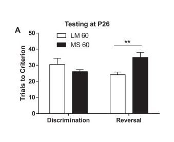
Early maternal separation impacts cognitive flexibility at the age of first independence in mice
Early life adversity is associated with increased risk for mental and physical health problems, including substance abuse. Changes in neural development caused by early life insults could cause or complicate these conditions. Maternal separation (MS) is a model of early adversity for rodents. Clear effects of MS have been shown on behavioral flexibility in rats,…