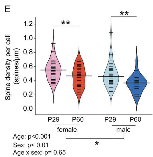
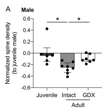
In humans, nonhuman primates, and rodents, the frontal cortices exhibit grey matter thinning and dendritic spine pruning that extends into adolescence. This maturation is believed to support higher cognition but may also confer psychiatric vulnerability during adolescence. Currently, little is known about how specific cell types in the frontal cortex mature or whether puberty plays a role in the maturation of some cell types but not others. Here, we used mice to characterize the spatial topography and adolescent development of cross-corticostriatal (cSTR) neurons that project through the corpus collosum to the dorsomedial striatum. We found that apical spine density on cSTR neurons in the medial prefrontal cortex decreased significantly between late juvenile (P29) and young adult time points (P60), with females exhibiting higher spine density than males at both ages. Adult males castrated prior to puberty onset had higher spine density compared to sham controls. Adult females ovariectomized before puberty onset showed greater variance in spine density measures on cSTR cells compared to controls, but their mean spine density did not significantly differ from sham controls. Our findings reveal that these cSTR neurons, a subtype of the broader class of intratelencephalic-type neurons, exhibit significant sex differences and suggest that spine pruning on cSTR neurons is regulated by puberty in male mice.
Kristen Delevich, Nana J Okada, Ameet Rahane, Zicheng Zhang, Christopher D Hall, Linda Wilbrecht, Sex and Pubertal Status Influence Dendritic Spine Density on Frontal Corticostriatal Projection Neurons in Mice, Cerebral Cortex, , bhz325, https://doi.org/10.1093/cercor/bhz325 (preprint available at https://www.biorxiv.org/content/biorxiv/early/2019/09/30/787408.full.pdf)