Tag: Joshua T Trachtenberg
-

Long-term, high-resolution imaging in the mouse neocortex through a chronic cranial window
To understand the cellular and circuit mechanisms of experience-dependent plasticity, neurons and their synapses need to be studied in the intact brain over extended periods of time. Two-photon excitation laser scanning microscopy (2PLSM), together with expression of fluorescent proteins, enables high-resolution imaging of neuronal structure in vivo. In this protocol we describe a chronic cranial…
-
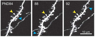
Transient and Persistent Dendritic Spines in the Neocortex In Vivo
Dendritic spines were imaged over days to months in the apical tufts of neocortical pyramidal neurons (layers 5 and 2/3) in vivo. A fraction of thin spines appeared and disappeared over a few days, while most thick spines persisted for months. In the somatosensory cortex, from postnatal day (PND) 16 to PND 25 spine retractions…