Category: Publications & Preprints
-
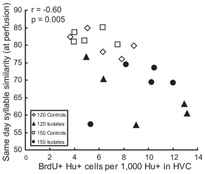
High levels of new neuron addition persist when the sensitive period for song learning is experimentally prolonged
Socially reared zebra finch males imitate a song they hear during posthatching days 30–65; during this time, many new neurons are added to the high vocal center (HVC), a forebrain nucleus necessary for the production of learned song. New neuron addition drops sharply after day 65, and no new songs are imitated. In contrast, male…
-
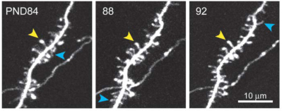
Spine growth precedes synapse formation in the adult neocortex in vivo
Dendritic spines appear and disappear in an experience-dependent manner. Although some new spines have been shown to contain synapses, little is known about the relationship between spine addition and synapse formation, the relative time course of these events, or whether they are coupled to de novo growth of axonal boutons. We imaged dendrites in barrel…
-

Experience-dependent and cell-type specific spine growth in the neocortex
Functional circuits in the adult neocortex adjust to novel sensory experience, but the underlying synaptic mechanisms remain unknown. Growth and retraction of dendritic spines with synapse formation and elimination could change brain circuits. In the apical tufts of layer 5B (L5B) pyramidal neurons in the mouse barrel cortex, a subset of dendritic spines appear and…
-
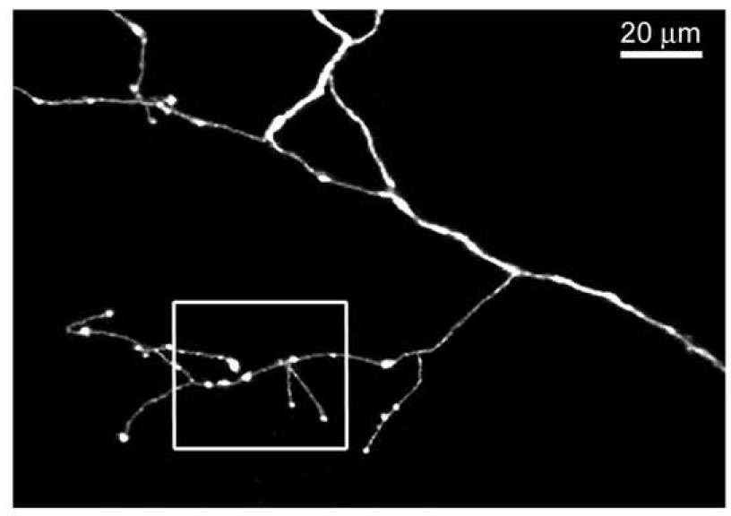
Cell type-specific structural plasticity of axonal branches and boutons in the adult neocortex
We imaged axons in layer (L) 1 of the mouse barrel cortex in vivo. Axons from thalamus and L2/3/5, or L6 pyramidal cells were identified based on their distinct morphologies. Their branching patterns and sizes were stable over times of months. However, axonal branches and boutons displayed cell type-specific rearrangements. Structural plasticity in thalamocortical afferents…
-
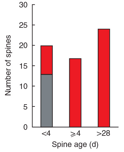
Transient and Persistent Dendritic Spines in the Neocortex In Vivo
Dendritic spines were imaged over days to months in the apical tufts of neocortical pyramidal neurons (layers 5 and 2/3) in vivo. A fraction of thin spines appeared and disappeared over a few days, while most thick spines persisted for months. In the somatosensory cortex, from postnatal day (PND) 16 to PND 25 spine retractions…