About webmatr0n

This author has not yet filled in any details.
So far webmatr0n has created 85 blog entries.

Choice suppression is achieved through opponent but not independent function of the striatal indirect pathway in mice

The dorsomedial striatum (DMS) plays a key role in action selection, but little is known about how direct and indirect pathway spiny projection neurons (dSPNs and iSPNs) contribute to choice suppression in freely moving animals. Here, we used pathway-specific chemogenetic manipulation during a serial choice foraging task to test opposing predictions for iSPN function generated by two theories: 1) the ‘select/suppress’ heuristic which suggests iSPN activity is required to suppress alternate choices and 2) the network-inspired Opponent Actor Learning model (OpAL) which proposes that the weighted difference of dSPN and iSPN activity determines choice. We found that chemogenetic activation, but not inhibition, of iSPNs disrupted learned suppression of nonrewarded choices, consistent with the predictions of the OpAL model. Our findings suggest that iSPNs’ role in stopping and freezing does not extend in a simple fashion to choice suppression. These data may provide insights critical for the successful design of interventions for addiction or other conditions in which suppression of behavior is desirable.

Kristen Delevich, Benjamin Hoshal, Anne GE Collins, Linda Wilbrecht, Choice suppression is achieved through opponent but not independent function of the striatal indirect pathway in mice, BioRxiv, https://www.biorxiv.org/content/10.1101/675850v3
doi: https://doi.org/10.1101/675850
 

Choice suppression is achieved through opponent but not independent function of the striatal indirect pathway in mice2022-06-18T20:52:41+00:00

Sex and Pubertal Status Influence Dendritic Spine Density on Frontal Corticostriatal Projection Neurons in Mice

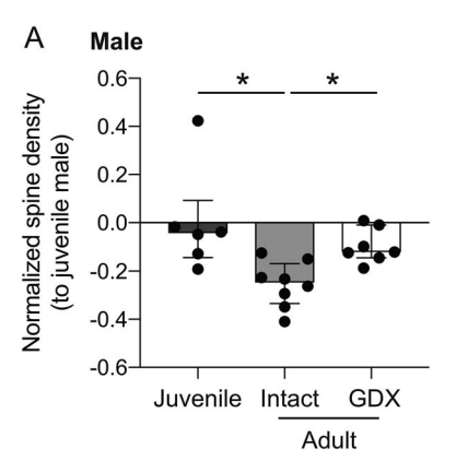
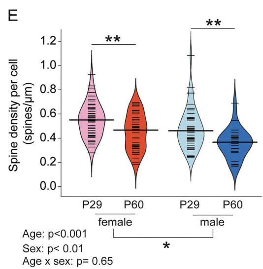
In humans, nonhuman primates, and rodents, the frontal cortices exhibit grey matter thinning and dendritic spine pruning that extends into adolescence. This maturation is believed to support higher cognition but may also confer psychiatric vulnerability during adolescence. Currently, little is known about how specific cell types in the frontal cortex mature or whether puberty plays a role in the maturation of some cell types but not others. Here, we used mice to characterize the spatial topography and adolescent development of cross-corticostriatal (cSTR) neurons that project through the corpus collosum to the dorsomedial striatum. We found that apical spine density on cSTR neurons in the medial prefrontal cortex decreased significantly between late juvenile (P29) and young adult time points (P60), with females exhibiting higher spine density than males at both ages. Adult males castrated prior to puberty onset had higher spine density compared to sham controls. Adult females ovariectomized before puberty onset showed greater variance in spine density measures on cSTR cells compared to controls, but their mean spine density did not significantly differ from sham controls. Our findings reveal that these cSTR neurons, a subtype of the broader class of intratelencephalic-type neurons, exhibit significant sex differences and suggest that spine pruning on cSTR neurons is regulated by puberty in male mice.

Kristen Delevich, Nana J Okada, Ameet Rahane, Zicheng Zhang, Christopher D Hall, Linda Wilbrecht, Sex and Pubertal Status Influence Dendritic Spine Density on Frontal Corticostriatal Projection Neurons in MiceCerebral Cortex, , bhz325, https://doi.org/10.1093/cercor/bhz325 (preprint available at https://www.biorxiv.org/content/biorxiv/early/2019/09/30/787408.full.pdf)

Sex and Pubertal Status Influence Dendritic Spine Density on Frontal Corticostriatal Projection Neurons in Mice2020-02-13T03:35:21+00:00

Distentangling the systems contributing to changes in learning during adolescence

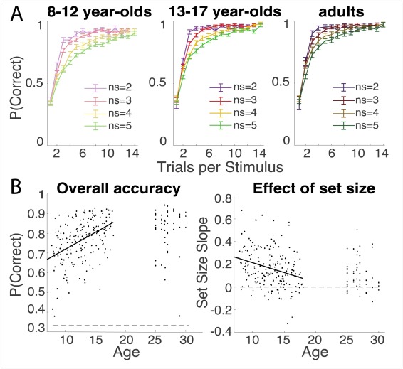
Multiple neurocognitive systems contribute simultaneously to learning. For example, dopamine and basal ganglia (BG) systems are thought to support reinforcement learning (RL) by incrementally updating the value of choices, while the prefrontal cortex (PFC) contributes different computations, such as actively maintaining precise information in working memory (WM). It is commonly thought that WM and PFC show more protracted development than RL and BG systems, yet their contributions are rarely assessed in tandem. Here, we used a simple learning task to test how RL and WM contribute to changes in learning across adolescence. We tested 187 subjects ages 8 to 17 and 53 adults (25-30). Participants learned stimulus-action associations from feedback; the learning load was varied to be within or exceed WM capacity. Participants age 8-12 learned slower than participants age 13-17, and were more sensitive to load. We used computational modeling to estimate subjects’ use of WM and RL processes. Surprisingly, we found more protracted changes in RL than WM during development. RL learning rate increased with age until age 18 and WM parameters showed more subtle, gender- and puberty-dependent changes early in adolescence. These results can inform education and intervention strategies based on the developmental science of learning.

Sarah L. Master, Maria K. Eckstein, Neta Gotlieb, Ronald Dahl, Linda Wilbrecht, Anne G.E. Collins, Distentangling the systems contributing to changes in learning during adolescence, Developmental Cognitive Neuroscience (2019),
https://doi.org/10.1016/j.dcn.2019.100732, http://www.sciencedirect.com/science/article/pii/S1878929319303196

Distentangling the systems contributing to changes in learning during adolescence2019-11-20T23:08:54+00:00

Variation in early life maternal care predicts later long range frontal cortex synapse development in mice

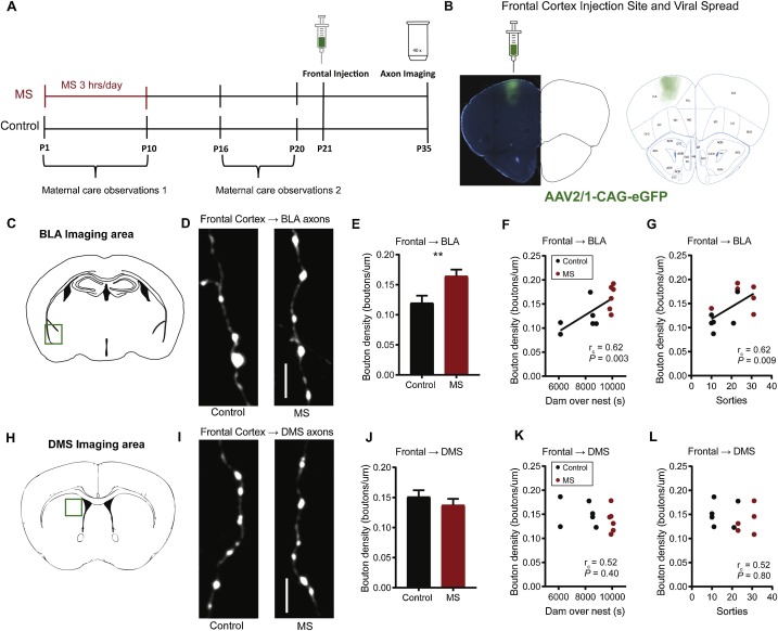
Empirical and theoretical work suggests that early postnatal experience may inform later developing synaptic connectivity to adapt the brain to its environment. We hypothesized that early maternal experience may program the development of synaptic density on long range frontal cortex projections. To test this idea, we used maternal separation (MS) to generate environmental variability and examined how MS affected 1) maternal care and 2) synapse density on virally-labeled long range axons of offspring reared in MS or control conditions. We found that MS and variation in maternal care predicted bouton density on dorsal frontal cortex axons that terminated in the basolateral amygdala (BLA) and dorsomedial striatum (DMS) with more, fragmented care associated with higher density. The effects of maternal care on these distinct axonal projections of the frontal cortex were manifest at different ages. Maternal care measures were correlated with frontal cortex → BLA bouton density at mid-adolescence postnatal (P) day 35 and frontal cortex → DMS bouton density in adulthood (P85). Meanwhile, we found no evidence that MS or maternal care affected bouton density on ascending orbitofrontal cortex (OFC) or BLA axons that terminated in the dorsal frontal cortices. Our data show that variation in early experience can alter development in a circuit-specific and age-dependent manner that may be relevant to early life adversity.

A. Wren Thomas, Kristen Delevich, Irene Chang, Linda Wilbrecht, Variation in early life maternal care predicts later long range frontal cortex synapse development in mice, Developmental Cognitive Neuroscience (2009)
https://doi.org/10.1016/j.dcn.2019.100737, http://www.sciencedirect.com/science/article/pii/S187892931930324X

Variation in early life maternal care predicts later long range frontal cortex synapse development in mice2019-11-20T23:02:33+00:00

Kristen Delevich Awarded Tourette Association of America Funding

The Tourette Association of America has awarded its Young Investigator Award to Kristen Delevich for her research project, Studying the Influence of Hormones on the Brain. This work seeks to understand the influence of puberty on brain circuits involved in behavioral control, in an effort to elucidate why Tourette symptoms typically change during adolescence. Congratulations, Dr. Delevich!

Kristen Delevich Awarded Tourette Association of America Funding2019-10-17T22:14:02+00:00

Coming of age in the animal kingdom

Coming of age in the animal kingdom2019-09-27T15:20:08+00:00

Imaging striatal dopamine release using a nongenetically encoded near infrared fluorescent catecholamine nanosensor

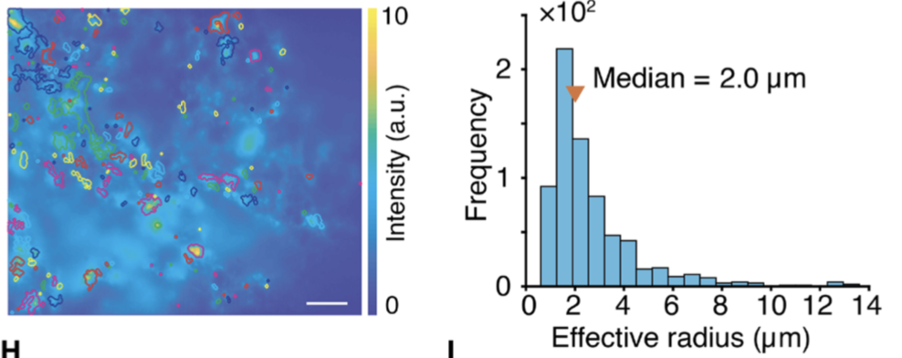
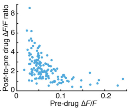
Neuromodulation plays a critical role in brain function in both health and disease, and new tools that capture neuromodulation with high spatial and temporal resolution are needed. Here, we introduce a synthetic catecholamine nanosensor with fluorescent emission in the near infrared range (1000–1300 nm), near infrared catecholamine nanosensor (nIRCat). We demonstrate that nIRCats can be used to measure electrically and optogenetically evoked dopamine release in brain tissue, revealing hotspots with a median size of 2 µm. We also demonstrated that nIRCats are compatible with dopamine pharmacology and show D2 autoreceptor modulation of evoked dopamine release, which varied as a function of initial release magnitude at different hotspots. Together, our data demonstrate that nIRCats and other nanosensors of this class can serve as versatile synthetic optical tools to monitor neuromodulatory neurotransmitter release with high spatial resolution.

Abraham G. Beyene, et al., Imaging striatal dopamine release using a nongenetically encoded near infrared fluorescent catecholamine nanosensor, 5(7) Science Advances eaaw3108 (2019)(local PDF).

Imaging striatal dopamine release using a nongenetically encoded near infrared fluorescent catecholamine nanosensor2019-07-17T19:51:56+00:00

New nIRCats Paper with the Landry Lab

We just published our first collaborative paper with the Landry lab in Science Advances. Beyene et al. shows that new nIRCats enable detection of dopamine release with high spatial resolution and are compatible with dopamine receptor pharmacology. There is brief coverage in Nature and a link to the paper is here.

New nIRCats Paper with the Landry Lab2019-07-17T19:39:50+00:00

Neuroscience: Sex Hormones at Work in the Neocortex

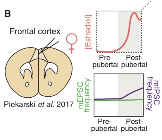
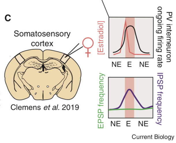
Sex hormones have easy access to the brain and their receptors are expressed by cortical neurons. Until recently, little was known about what impact, if any, they have on cortical processing. New data reveal that estradiol potently alters inhibitory neurotransmission in the neocortex.

Kristen Delevich, David Piekarski, Linda Wilbrecht, Neuroscience: Sex Hormones at Work in the Neocortex,
29(4) Current Biology R122–R125 (2019) https://doi.org/10.1016/j.cub.2019.01.013, http://www.sciencedirect.com/science/article/pii/S0960982219300156

Neuroscience: Sex Hormones at Work in the Neocortex2019-02-22T15:49:13+00:00

Adolescence and “Late Blooming” Synapses of the Prefrontal Cortex

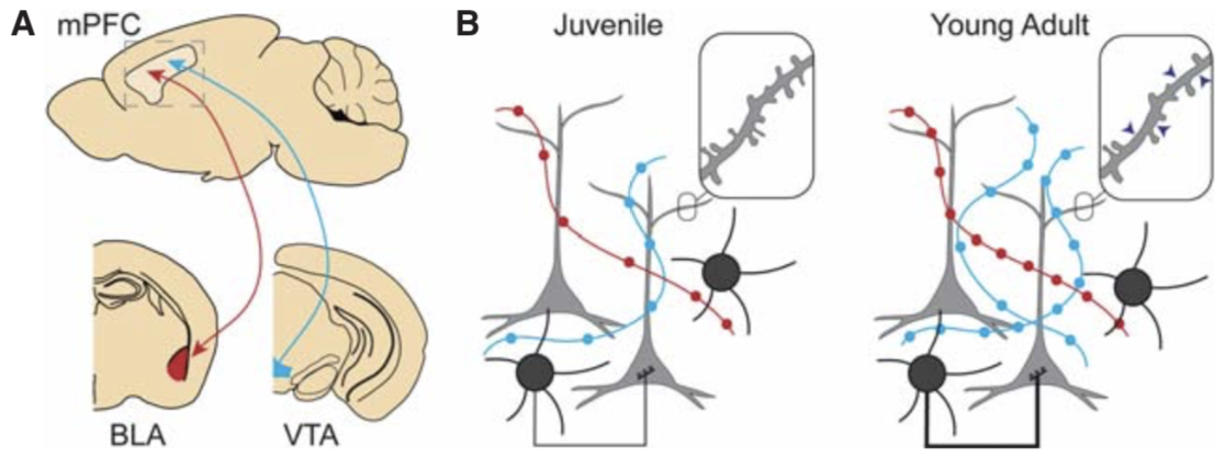
The maturation of the prefrontal cortex (PFC) during adolescence is thought to be important for cognitive and affective development and mental health risk. Whereas many summaries of adolescent development have focused on dendritic spine pruning and gray matter thinning in the PFC during adolescence, we highlight recent rodent data from our laboratory and others to call attention to continued synapse formation and plasticity in the adolescent period in specific cell types and circuits. In particular, we highlight changes in inhibitory neurotransmission onto intratelencephalic (IT-type) projecting cortical neurons and late expansion of connectivity between the amygdala and PFC and the ventral tegmental area and PFC. Continued work on these “late blooming” synapses in specific cell types and circuits, and their interrelationships, will illuminate new opportunities for understanding and shaping the biology of adolescent development. We also address which aspects of adolescent PFC development are dependent on pubertal processes.

Kristen Delevich, A. Wren Thomas, and Linda E. Wilbrecht, Adolescence and “Late Blooming” Synapses of the Prefrontal Cortex [local pdf], Cold Spring Harb Symp Quant Biol (2019)

Adolescence and “Late Blooming” Synapses of the Prefrontal Cortex2019-02-01T12:32:47+00:00